File:Schizophrenia PET scan.jpg
Jump to navigation
Jump to search
No higher resolution available.
Schizophrenia_PET_scan.jpg (224 × 248 pixels, file size: 23 KB, MIME type: image/jpeg)
Summary
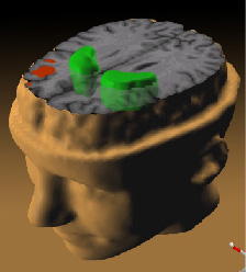
illustration of Schizophrenia's effect on the brain; taken from here archive copy at the Wayback Machine
- Source: Andreas Meyer-Lindenberg, M.D., Ph.D., NIMH Clinical Brain Disorders Branch on the tree of life.
While patients performed a working memory task, the less the prefrontal cortex (red) activated, the more dopamine increased in the striatum (green).
Abstract of study is here.
Licensing
This image is a work of the National Institutes of Health, part of the United States Department of Health and Human Services, taken or made as part of an employee's official duties. As a work of the U.S. federal government, the image is in the public domain.
 |
||
| This file has been identified as being free of known restrictions under copyright law, including all related and neighboring rights. | ||
File history
Click on a date/time to view the file as it appeared at that time.
| Date/Time | Thumbnail | Dimensions | User | Comment | |
|---|---|---|---|---|---|
| current | 12:07, 30 November 2005 | | 224 × 248 (23 KB) | wikimediacommons>Skagedal | illustration of Schizophrenia's effect on the brain; taken [http://www.nih.gov/news/pr/jan2002/nimh-28.htm from here] *Source: Andreas Meyer-Lindenberg, M.D., Ph.D., NIMH Clinical Brain Disorders Branch ''While patients performed a working memory t |
File usage
The following page uses this file: